
Archives
High-tech tool helps students learn about TMJ
By MARY COCHRANE
Contributing Editor
In an unusual collaboration, a professor in the School of Dental Medicine has joined forces with faculty members in the School of Architecture and Planning to develop a high-tech teaching tool to assist in the education of dental students and patients.
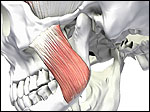
A high-tech teaching tool developed by UB faculty members is helping dental students understand the TMJ.
Lida Radfar, assistant professor in the dental school, needed to make the most of the few hours she has in a clinical procedures course (CLD 822) to give students a proper introduction to a key point of anatomy.
So she called a friend in the School of Architecture and Planning for help. That friend—Shahin Vassigh, associate professor of architecture—in turn enlisted the expertise of her colleague, Omar Khan, assistant professor of architecture, and a highly innovative teaching tool was born.
Vassigh and Khan are co-directors of the Center for Virtual Architecture, which is involved in the investigation and application of digital technology in architectural research, design and education. With a grant from the Educational Technology Center (ETC), the trio collaborated to create a visual model of the temporomandibular joint, or TMJ. Carole Ann Fabian, ETC director, said the center's annual awards are supported by the Office of the Chief Information Officer and administered through the ETC.
The TMJ is a bilateral, biconcave joint that is positioned between the mandible and the temporal bone. Although the major function of the TMJ is to open, close and approximate movement of the jaws during mastication of food, other assisting functions include swallowing, phonation, yawning and suckling. Jaw movement is a complex, multidimensional process; the joints are passive and the muscles are active structures.
"I have a very limited time with these students—three hours—and TMJ is a complex area," Radfar said. "So I want them to see something, to have a picture in their minds. They need something to look at so they can understand it."
UB has a Temporomandibular Disorders Clinic and dental students also will learn about TMJ in later classes from the specialists here at the university. But Radfar said she wants her clinical procedures students to understand this important area of dentistry right from the start.
"With its complex anatomy and three-dimensional operation, TMJ is a subject that remains unclear and incompletely understood on first encounter," Radfar said. "Misunderstanding the multifunctional TMJ may lead to clinically misdiagnosed situations and eventually untreated or maltreated patients."
The three professors set out to create a tool that will result in students' better understanding the functional anatomy of TMJ, and subsequently perform better examinations and better clinical diagnoses of patient conditions.
The instructional project includes high-quality digital graphics, realistic three-dimensional digital models, animation and audio narration to show the functional anatomy of TMJ.
"The visual is helpful because it shows students how the joint and muscle of the jaw move together," Radfar said. "They see when things are moving, they are moving together. This is going to help them understand this complex structure."
Radfar provided the textual content for the project, as well as the human skull that was used for demonstration of the joint and its related muscles. Khan provided the interface design for delivering the entire content of the module. The project can be distributed in CD-rom, DVD or Web formats.
Vassigh and Khan used many of the same techniques, technologies and approaches in the TMJ project that are used in an instructional software program called "Interactive Structures: Visualizing Structural Behavior," which was funded by the U.S. Department of Education. Vassigh developed and used that teaching tool in two courses in structural science in the School of Architecture and Planning.
The TMJ project is Vassigh's first collaboration with an unrelated field. "The modeling of a skull and providing an interface for the students to access the information visually was architecture's contribution to the project," Vassigh said.
The team overlaid the skull with "an intense mesh of points," which they then scanned into a computer modeling program called Rhino, enabling them to provide a realistic, three-dimensional model of the skull, Vassigh said.
After adding modeled muscles, they created a series of animations to show the anatomy of the joint and its surrounding system. They then compiled all information into a computer-authoring program, which provided the interface. The program was then copied onto a CD so it can be used in the classroom, as well as distributed to students for individual study.
The team plans to seek external grant money to expand the scope of the project, Vassigh said. Radfar said she would like to create similar teaching tools for classes in her areas of specialty—salivary glands and oral lesions—as well as for individuals treated in UB's salivary gland dysfunction clinic, which welcomes patients from throughout Western New York.